Wednesday, October 28, 2015

Diffusion Osmosis Lab - Excercise 1D


Claculation of Water Potential from Experimental Data 

1. The solute potential can be calculated using the following formula:


2. Knowing the solute potential of the solution and knowing  that the pressure potential of the solution is zero, allows you to calculate the water potential of the solution. The water potential will be equal to the solute potential of the solution.


Conclucsion/Analysis:


Calculation the water potential will be useful when predicting the movement of water when it comes to different solutions of varying concentrations. It was observed from the potato cores that water will always move from areas of high concentration to areas of low concentration. The water of the potato cores left the potato cores and went instead into the sucrose solution. This trend was followed as we observed the percent change in the potato cores have a direct relationship with the molarity of the sucrose solution, meaning that as the sucrose solution molarity increased, so did the percent change of the potatoe cores.

Diffusion Osmosis Lab Part 1B

Part 1B:
Introduction / Purpose:


The purpose of this experiment was to explore the relationship between the solute concentration and the passage of water through a selectively permeable membrane by the natural process of osmosis. The solute concentration may be isotonic, hypotonic, or hypertonic. Isotonic solutions will have the same amount of solute concentration. There will be no net change in the amount of water in each solution. The two solutions are said to be hypertonic if there is more solute concentration inside of the cell than there is outside. A solution is said to be hypotonic when there is less solute concentration inside of the cell. For this experiment, a hypertonic solution was placed in a dialysis tubing bag and let to sit in a hypotonic solution for 30 minutes.


Procedure:


1.  Obtain six 30-cm strips of presoaked in water dialysis tubing.


2. Tie a knot in one end of each piece of dialysis tubing to form 6 bags. Pour approximately 15-25 mL of each of the following solutions into separate bags; (distilled water, 0.2M sucrose, 0.4M sucrose, 0.6M sucrose, 0.8M sucrose, 1.0M sucrose) Leave sufficient space for the expansion of the contents in the bag.


3. Rinse each bag gently with distilled water to remove any sucrose spilled during the filling.


4. Carefully blot the outside of each bag and record in Table 1.2 the initial mass of each bag, expressed in grams.


5. Place each bag in an empty 250-mL cup and label the beaker to indicate the molarity of the solution in the dialysis bag.


6. Now fill each beaker two-thirds full with distilled water. Be sure to completely submerge each bag.


7. Let them stand for 30 minutes.


8. At the end of 30 minutes remove the bags from the water. Carefully blot and determine the mass of each bag.


9. Record your group's data in Table 1.2 and fill Graph 1.1.


Conclusion/Analysis:


From the experiment, it is possible to conclude that when the bags were placed in a hypotonic solution, meaning that the solute concentration inside of the bag was greater than the solute concentration outside of the bag, the bags’ masses increased, as shown in Table 1.2. This may be due to water moving to the inside of the cell due to diffusion, water’s tendency to move from areas of high concentration to areas of low concentration. In the case of hypotonic solutions, water must rush into the bag since there is an increasingly less amount of water in each bag as the molarity increases. This would explain why bags with a higher molarity would experience a greater change in their mass, as noted in Table 1.2. Our graph may be skewed, however, due to human error, whether it be in performing the procedure or doing the calculations.

Tuesday, October 27, 2015

Diffusion Osmosis Lab Part 1C

Introduction:


Water potential is a measurement of how likely it is that water will leave on place in order to go another. When in an isotonic solution, water potential will be lower since there will be no net movement of water. Water potential is dependent on the pressure potential and the solute potential. Adding both of these will yield the water potential. An increase in the solute potential will lower the water potential, while an increase in pressure potential will increase the water potential. In this experiment, potato cores and sucrose solutions, all of varying molar concentration, were used in order to determine the water potential of potato cores.


Procedure:


1. Pour 100 mL of the assigned solution into a labeled 250-mL beaker. Slice a potato into discs that are approximately 3 cm thick.


2. Use a cork borer (approximately 5 mm in inner diameter) to cut four potato cylinders. Do not include any skin on the cylinders. You need four potato cylinders for each beaker.


3. Keep your potato cylinders in covered beaker until it is your turn to use the balance.


4. Determine the mass of the four cylinders together and record the mass in Table 1.4. Put the four cylinders into the beaker of sucrose solution.


5. Cover the beaker with plastic wrap to prevent evaporation.


6. Let it stand overnight.


7. Remove the cores from the beakers, blot them gently on a paper towel, and determine their total mass.


8. Record the final mass in Table 1.4 and record the class data in Table 1.5. Calculate the percent change as you did in Exercise 1B. Do this for both your individual results and the class average.


9. Graph both your individual data and the class average for the percent change in mass in Table 1.4.


Conclusion/Analysis:


From this experiment it was possible to determine that the water potential of the potato cells was higher than that of the sucrose solution. After the 48 hours of having the potato cells be in the sucrose solution had passed, the potato cores were soggy and softer. It is possible that these observations were due to the loss of water from the cells, therefore indicating that the water moved from an area of high water potential to that of a lower one. For this reason, one can then determine that the water potential inside of the cells of the potato cores was higher than the water potential in the sucrose solution.
In order for the cells to reach an isotonic state, our individual group data showed that the net movement of water was zero when the potato cores were immersed in .3 M solution. This can found by analyzing Graph 1.2 and seeing that an isotonic state is when the potato cores are submerged in a .3M solution, since there is no net movement of water and no change in the potato core mass. Our data slightly differs from the class data. When analyzing the data from class in Graph 1.2, one can see that an isotonic state for the potato core cells is reached the potato cores are put in around a roughly 4.7M solution.


Diffusion Osmosis Lab Part 1A

Part 1A:
Introduction / Purpose: 

The purpose of this experiment was to demonstrate the concept of selective permeability. When a membrane is selectively permeable, this means that only certain molecules are allowed to pass through. Generally, smaller molecules are able to pass through the membrane with ease, but larger molecules take a longer amount of time, or cannot pass through period. The process by which molecules pass through the membrane is called dialysis. In our experiment we measured to see how selectively permeable the dialysis tubing  is. We filled the dialysis tubing up with a 15% Glucose / 1% Starch solution and let it sit in a cup of water filled with H2O and Iodine. After 30 minutes it was clear that Iodine molecules had passed into the membrane and glucose had escaped.  

Procedure:

1. Obtain a 30-cm piece of 2.5-cm dialysis tubing that has been soaking in water. Tie off one end of the tubing to form a bag.

2. Test the 15% Glucose / 1% Starch solution for the presence of glucose. Your teacher may have you do a Benedict's test or a Cit. Record the results in Table 1.1.


3. Place 15 mL of the 15% Glucose / 1% Starch solution in the bag. Tie off the other end of the bag, leaving sufficient space for the expansion of the contents in the bag. Record the color of the solution in Table 1.1.


4. Fill a 250 mL cup two-thirds full with distilled water. Add approximately 4 mL of Lugol's solution to the distilled water and record the color of the solution in Table 1.1. Test this solution for glucose and record the results in Table 1.1.

5. Immerse the bag in the beaker of solution.


6. Allow your setup tp stand for approximately 30 minutes or until you see a distinct color change in the bag or the beaker. Record the final color of the solution in the bag, and of the solution in the beaker, in Table 1.1.


7. Test the liquid in the beaker and in the bag for the presence of glucose. Record the results in Table 1.1.


Conclusion/Analysis:

Based on the results of the experiment, it is possible to conclude that the dialysis tubing was selectively permeable. Before being immersed in the iodine, when tested, the dialysis bag showed presence of glucose. This was demonstrated with the paper of the Clinistix turning a dark brown color, signaling glucose. On the other hand, there was no glucose presence detected by the Clinistix when dipped into the iodine, since the stick turned mint-green, the color for no glucose. However, when the bag and the iodine were tested a second time for the presence of glucose, after the bag had sat in the iodine for 30 minutes, the Clinistix strip turned lime-green for both the bag and the iodine. This told us that the glucose molecules of the 15% glucose / 1% starch solution were small enough to go through the membrane of the dialysis tubing and mix in with the iodine. However, the lack of starch indicates that these starch molecules were too large in order to be able to pass through the dialysis tubing membrane. Evidence of the lack of starch is the fact that iodine will turn the starch a dark blue color when the two substances are mixed together. However, at the end of the experiment, the solution in the cup remained red, its original color, but the dialysis bag was now a dark blue color. This indicated that the starch molecules had remained inside of the bag and not drained into the cup. It was instead the iodine molecules which were able to pass through the dialysis tubing membrane and mix with the starch molecules inside of the bag, thus turning the bag a dark blue color.

Chapter 8 Portfolio

  • Compare and contrast Endergonic and Exergonic reactions - include a model of each showing the change in free energy from reactants and products.

Endergonic type of reactions absorb energy. They are anabolic and non-spontaneous, meaning that they require energy input. They will also have a positive net change in free energy. Exergonic reactions will release energy into the surrounding area. They are catabolic and spontaneous, meaning that they do not require energy input. Their net change in free energy is negative. This free energy is able to perform work, as long as the temperature and pressure are maintained constant.


Source: www.slideshare.net


  • Explain the key role of ATP in energy coupling reactions. Provide a model showing energy before and after phosphorylation of the reactants.


One ATP, also known as Adenosine triphosphate, molecule  is composed of:


  • 1 Ribose, sugar, molecule
  • 1 Adenine, nitrogenous base, molecule
  • 3 Phosphate groups


Energy couple is when an exergonic reaction is used to drive an endergonic one. Through hydrolysis,  one of the three phosphate groups will break off. The energy from this process comes from the change to a state of lower free energy. It DOES NOT come from the phosphate group breaking off. Phosphorylation is when the phosphate group that broke of the original ATP molecule binds to another new molecule, which is referred to as the phosphorylated intermediate. This then in a sense regenerates the ADP molecule into ATP.


Source: cnx.org

  • Analyze data showing how changes in enzyme structure, substrate concentration, and environmental conditions (pH, temperature, salinity, etc.) affect enzymatic activity - ie. include a graph of each example and predict the effects when one of the parameters is further changed. Justify and explain your predictions.


Enzymes will function best only in certain conditions. Changes in the temperature and the pH of the substrate may cause the enzyme to become inefficient. The two graphs below demonstrate the effects of changes of pH on the enzymes pepsin and trypsin. The enzyme’s rate of reaction will peak only at either a pH level of 2 or a pH level of 8. As either the pHs stray from the optimal pH, whether there is an increase or a decrease in pH, the rate of reaction goes down.
The same thing is demonstrated in the second graph. The enzyme’s highest rate of reaction will only occur at a specific temperature.


Source: Pearson Education Inc. 

  • Describe the relationship between the structure and function of enzymes.


Enzymes are a special type of protein, relatively large in size, that will speed up a chemical reaction. They are able to do this without change to the amount of free energy available. This makes enzymes catalysts. The substance that a catalysts, in this case enzymes,  acts on is referred to as a substrate. Only specific enzymes may catalyze certain molecules. What molecules the enzyme may act on is dependent on the enzyme's structure. The substrate must be able to bond to the enzyme's activation site and from the enzyme-substrate complex. It is only after this that the speeding up of the reaction occurs.  

  • Explain how enzymes accomplish biological catalysis.  Provide examples.


Enzyme’s serve to speed up metabolic reactions without the use of free energy. Enzymes are able to achieve this by. however, by LOWERING the amount of free energy REQUIRED to make reactants into the necessary products. Without enzyme’s product production would become inefficient since the reactions would take too long.
Source: www.slideshare.net

  • Describe how enzyme-mediated reactions can be controlled through competitive and noncompetitive interactions.
Similar to the definition of inhibit, which is to hinder or prevent, and inhibitor will bond with an enzyme in order to stop its activity. Inhibitors can either be competitive or noncompetitive.
Competitive inhibitors will in a sense “compete” with the substrate. They will bind to the active site of an enzyme and compete with the substrate for the site and try to block the substrate from binding. On the other hand, non-competitive inhibitors will take a different route to stop the reaction and will instead bind to a different section of the enzyme. They will then change the shape of enzyme, thus making the active site less effective and stopping the reaction. Inhibitors control enzyme-mediated reactions by stopping them.


  • Propose experimental designs by which the rate of enzyme function can be measured and studied.


A useful way to measure the functionality of an enzyme would be to measure the time it takes for the enzyme to actually complete the process, or in other words turn the substrate into products. This experiment would be similar to the one recently conducted in class. One would have to do several timed trials to see how much of the substrate reacts. Time would being when the catalyst is added to the substrate. The time would stop when the inhibitor is added. The, using a titration system, one would measure the amount of substrate still present or the amount that reacted, and thus measure the functionality of the enzyme. Enzymes that work better would cause more reaction in a shorter span of time.

Sunday, October 18, 2015

Chapter 7 Portfolio

  • Describe passive transport and explain its role in cellular systems


Passive transport is when materials are able to cross the cell membrane without the use of any energy in the form of ATP. Since the plasma membrane is selectively permeable, this means that only certain substances will be able to pass through. The membranes composition of fatty acid tails allows for nonpolar molecules to easily pass, without the use of a transport protein. It is through the process of diffusion that these molecules are able to do this.


  • Explain how membrane proteins play a role in facilitated diffusion of charged and polar molecules in general and in relation to the specific molecules below.


SInce part of the plasma membrane is nonpolar, polar molecules will require special proteins that will carry them across the cell membrane when the cell needs them.
    • Glucose transport
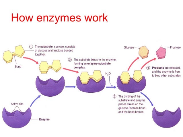
This protein is used to transport glucose molecules across the membrane. Located on the membrane of red blood cells, this protein will only accept glucose molecules to bring them into the cell. This type of transport DOES NOT require ATP.

Source: greatcourse.cnu.edu.cn

    • Na+/K+ transport
Also known as the sodium-potassium pump, it is a type of active transport system, meaning that it does require ATP.  This protein transport sodium and potassium against their concentration gradient. This means that it will take sodium from areas of low concentration and take them to areas of high concentrations. This helps the cell maintain concentration gradients that differ from their environment. It is like pushing a boulder up a hill instead of letting it roll down.
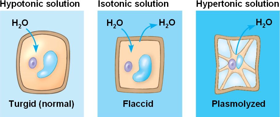

Source: www.vi.cl


  • Explain the terms: hypotonic, hypertonic or isotonic in relationship to the internal environments of cells. Illustrate each environment with a model and explain which environment is best for plant and animal cells. Justify and explain your model with evidence.


Hypotonic:
This is when there is less solute concentration inside of the cell. Water will rush INTO the cell. If immersed in a hypotonic environment, cell will SWELL and possibly burst (also known as a lysed cell) However, plant cells will not burst due to their cell walls. Is actually the best environment for plant cells.


Hypertonic:
This is when there is more solute concentration inside of the cell than there is outside. Water will rush OUT of the cell. If immersed in a hypertonic solution, the cell will shrivel.


Isotonic:
This is when the solute concentration is the same inside and outside of the cell. Animal cells, which do not have a cell wall will want to be in an isotonic environment.
Source: garrickray.weebly.com


  • Describe active transport. Propose a model illustrating this process.


Active transport is when ATP is necessary in order to get materials across the plasma membrane. This type of transport mainly takes place in the proteins that are embedded into the plasma membrane, including the sodium potassium pump. There also exists the electrogenic pump, which serves to transport protons across the membrane.

Source: bioap.wikispaces.com


  • Describe the processes of endocytosis and exocytosis. Propose a model illustrating each process.

Endocytosis (Materials IN):
This process is used to bring materials INTO the cell. It may help to think of this process as “the cell eating,” also known as phagocytosis. The cell does this by creating vesicles from the plasma membrane to engulf particles from the extracellular fluid.


Exocytosis (Materials OUT):
The opposite of endocytosis, this process is when the cell transports materials out of it. It does this by creating transport vesicles inside of the cell. These vesicles will then travel to the membrane, fuse with it, and then release their contents. This is used to get rid of wastes and also export products.
Source: www.lrn.org


  • Create a visual representation/model (ie. graph or diagram) to make predictions about the exchange of molecules between an organism and its environment, the use of these molecules (ie. CHNOPS and incorporation into carbohydrates, proteins, lipids, nucleic acids, membrane structure, genetic information, etc.), and consequences to the organism if these molecules cannot be obtained.

The interaction between plant cells and their exchange with water is dependent on the type of solution they are immersed in. Plant cells will desire to be in is hypotonic solution, so that water may rush into them and keep the cell walls rigid and stiff. Isotonic environments will be detrimental to plant cells since there is no water going in or out. Hypertonic environments are worse, causing water to leave the plant cells, making the plant limp and soggy.


Souce: bio1151.nicerweb.com


Tuesday, October 13, 2015

Chapter 6 Portfolio

  • Compare and contrast the structure of Prokaryotic and Eukaryotic cells - be sure to include a discussion regarding the cellular organization of each.


There are several main differences between Prokaryotic and Eukaryotic cells. Overall, Eukaryotic cells will be more complex, larger, and found in animals, while prokaryotic cells will be simpler, smaller, and usually be unicellular organisms. However, they are rarely multicellular as well.


Similarities between the two types of cells are:
  • both have a plasma membrane
  • both contain cytosol and cytoplasm
  • both contain DNA (so that they may divide)
  • both contain ribosomes ( in order to create proteins) - However, ribosomes will be larger in Eukaryotic cells
  • both have vacuoles
  • both have a flagella


However, there are some major distinctions between the two, these being:


  • Prokaryotic cells LACK a membrane bound nucleus. Instead, the DNA is spread out throughout the cell in the cytoplasm.
  • Prokaryotic cells lack the following organelles:
    • Nucelous
    • Lysosomes
    • Peroxisomes
    • Microtubules
    • Endoplasmic Reticulum
    • Mitochondria (Prokaryotic cells actually have no membrane bound organelles)
    • Golgi Apparatus
    • Chloroplast (However, prokaryotic cells DO contain chlorophyll. It is instead scattered throughout the cytoplasm)


  • Cells walls of prokaryotic cells will be much more complex than those of eukaryotic cells.
  • Prokaryotic cells will only have one chromosome, while eukaryotic cells have several.


Sources:


  • http://www.diffen.com/difference/Eukaryotic_Cell_vs_Prokaryotic_Cell
  • http://www.shmoop.com/biology-cells/prokaryotic-cells.html


  • Describe the basic structure and functions of key cell organelles (nucleus, Golgi, ER, mitochondria, chloroplast, vacuoles, plasma membrane). Include a diagram/drawing of each organelle!


  • Nucleus


The nucleus plays a role in storing the genetic information for the cell and serves as the command center for the cell. The nucleus has double membranes and is continuous with the ER. It is constructed of chromatin (web-like and holds DNA) and also contains the nucleolus, which has no membrane. Here rRNA, used to create ribosomes, is made.
Source: www.ck12.org


  • Plasma Membrane


The plasma membrane is what surrounds the cell and encloses all of its contents. It is selectively permeable, meaning that only certain substances may go through. It is composed of phospholipids. These phospholipids have a polar phosphate head and non-polar lipid tails.


Source: www.buzzle.com


  • Endoplasmic Reticulum


It is composed of a phospholipid bilayer and helps the cell to carry around nutrients. There are two types:


  • The Rough ER has ribosomes attached to it. It aids in the synthesis and packaging of proteins. The ribosome will build amino acid chains, which will eventually become proteins.


  • The Smooth ER is in charge of storing enzymes which help to create lipids. These enzymes also help to make certain substances not toxic to the body. Also stores ions.


Source: micro.magnet.fsu.edu


  • Golgi Apparatus


The Golgi Apparatus is in charge of processing and packaging proteins so that they may be sent to wherever in the cell that they need to go. The golgi is made up of flattened membrane sacs called cisternae. In addition to being part of processing proteins, the golgi is also capable of creating macromolecules, including polysaccharides. The golgi’s cisternae are essential divided into two sides. The cis (meaning on the same side) is connected to the ER and transport vesicles will transport materials from the ER to the golgi. The other side is called the trans (meaning on the opposite side) and has vesicles that pinch off and travel to other sites. It is while products of the ER travel from the cis to trans side that they are modified.


Source: www.britannica.com


  • Chloroplasts


Chloroplasts are composed of interconnected sacs called thylakoids. Stacks of thylakoids are called granums. Chloroplast DNA, ribosomes, and enzymes are found in the fluid outside the thylakoids, known as stroma. Therefore the three spaces of chloroplasts are the thylakoid space, stroma space, and intermembrane space. Chloroplasts main job is to use the sun's energy during photosynthesis. By using the sun’s energy, chloroplasts are able to create glucose and oxygen from carbon dioxide and water.


Source: www.ducksters.com


  • Mitochondria


Mitochondria is surrounded by double membranes. The exterior membrane is smooth However, the inner membrane has infoldings called cristae, giving the inner membrane a larger surface area and increasing cellular respiration productivity. The organelle is divided into two spaces - the intermembrane space and the second is the mitochondrial matrix. This matrix contains the not only the mitochondrial DNA and ribosomes, but also enzymes necessary to catalyze steps of cellular respiration, the main function of mitochondria. During cellular respiration, mitochondria will convert glucose and oxygen into carbon dioxide and water, but also energy in the form of ATP. The enzyme necessary for ATP production is embedded into the inner membrane.


Source: blog.phoreveryoung.com


  • Vacuoles


Vacuoles help to give mainly plant cells structure. As plants are able to absorb more water, the water vacuole will grow large within the cell and causing pressures within the cell and giving plants their crisp look. When plants dry, the central vacuole does not have water, there is no pressure, and plants wilt. Other types of vacuoles can store a variety of things, including food, reserves of important compounds like proteins, pigments, and even poisonous compounds to protect plants against herbivores.


Source: Crash Course Biology on Youtube 


  • Explain how several internal membrane-bound organelles and other structural features (e.g., ER, ribosomes) work together to provide a specific function for the cell (e.g., synthesis of protein for export) and contribute to efficiency (e.g., increasing surface area for reactions, localization of processes).


Proteins are essential to life. However, there is a process to produce and ship proteins. The process begins in the nucleus. Here, DNA is stored. This DNA is coding for the construction of proteins. THe nucleus will secret ribosomes, which will assemble the proteins by assembling amino acids into polypeptides. These ribosomes may either be floating in the cell or they may be attached to the rough ER. The rough ER is connected to the golgi apparatus, which will then process and package these proteins in vesicles, which have phospholipid walls. These vesicles will then travel to parts of the cell or outside of the cell.


  • Explain the structure and function relationships between chloroplasts and mitochondria


Chloroplasts and mitochondria both work to help cells create the energy necessary for molecular functions. Chloroplasts are in charge of using the sun’s energy to convert carbon dioxide and water into glucose. Mitochondria then use this glucose to create energy in the form of ATP during the process known as cellular respiration. Chloroplasts are found only in plant cells. They contain thylakoids, which look like stacked poker chips. Both organelles have double membranes. The mitochondria's inner membrane has infoldings known as cristae. Within the inner membrane are is the mitochondrial matrix, which contains enzymes that will catalyze the steps of cellular respiration.


  • Relate structural and functional evidence in chloroplasts and mitochondria to the endosymbiotic theory of their origins.


This theory suggests that originally the chloroplasts and mitochondria were cells of their own, living outside of the eukaryotic cell. This theory is supported by the fact that both organelles can create energy and the fact that they are autonomous. They divide on their own and each have their own DNA, separate from the DNA of the rest of the cell.The theory is also supported by the fact that both the chloroplasts and mitochondria have membranes of their own within the cell, suggesting that they were originally enveloped by the cell, and kept instead of digested when they served the purpose of providing the cell with energy.


  • Represent graphically and explain the relationship between surface area to volume ratios as it relates to the efficiency of cellular work.

The surface area and volume of a cell are very important to its function. In order to work bests, cells will want to 
have a large surface area to volume ratio. As a cell grows, the SA:V decreases since volume increases at a faster pace than SA. Having a large surface area is beneficial since there is more area for the cell to dispose of waste and take in nutrients. A smaller cell volume is desirable because that means that once inside the cell, nutrients will have to travel less distance to get to where the cell needs them to be.


Source: www.tiem.utk.edu