- Compare the major features of chemoheterotrophic and photoautotrophic nutritional processes.
Chemoheterotrophic processes:
These processes occur in organisms that cannot create their own food, and instead must ingest a combination of proteins, lipids, and carbohydrates to create their food. It is the equivalent of cellular respiration. Organisms that depend on chemoheterotrophic processes will obtain their energy by consuming other organisms.
Photoautotrophic processes:
If an organism is dependent upon photoautotrophic processes, it will obtain its energy from primarily the sunlight and the carbon dioxide in the air. These organisms are primarily plants and will perform processes such as photosynthesis to convert sunlight, CO2, and water into sugar necessary to carry out cellular functions.
- Explain the inputs, major processes, and outputs of glycolysis, fermentation, and aerobic cellular respiration.
Glycolysis:
During glycolysis,a glucose molecule will be broken down to create two pyruvate molecules, which will then go on to become acetyl CoA before entering the Citric Acid Cycle. The breakdown occurs in two phases. During the first phase, the Energy Investment phase, 2 molecules of ATP are consumed and turned into 2 ADP molecules and 2 Phosphate molecules.
In the second stage, the Energy Payoff phase, 4 ATP molecules are formed and NAD+ is reduced into 2 NADH and 2 H+, by the addition of 4 electrons and 4 H+. 2 Pyruvate and 2 H2O molecules are also formed.
 | |
|
Fermentation:
Fermentation will occur in the case that there is no oxygen readily available. For this reason it is considered an anaerobic process. Fermentation will use substrate-level phosphorylation to generate ATP and regenerate NAD+, the electron acceptor in glycolysis.
The two types of fermentation that occur are alcohol and Lactic Acid fermentation. In alcohol fermentation, pyruvate is converted into ethanol and CO2 is released. NADH is also regenerated into NAD+. Alcohol fermentation usually deals with yeast and bacteria.
In Lactic Acid fermentation, pyruvate is reduced into NADH, instead of ethanol. Lactate is a by product. However, no CO2 is released. This process occurs mainly in animals and is the cause for muscle fatigue. Yet, only 2 ATP molecules will be produced instead of the usual 38 ATP created during cellular respiration.
Aerobic Cellular Respiration:
For this process to occur it is necessary that oxygen be present. There are three phases in cellular respiration. The first phase is glycolysis. The input for this phase is 1 glucose molecule. The output of the process will be 2 molecules of pyruvate. These molecules will then be transported from the cytosol into the mitochondrial matrix by transport proteins to be oxidized into acetyl CoA. From there, the Citric Acid cycle will occur. Since each cycle requires only 1 molecule of CoA, the cycle will run twice. After the cycle has run twice, the net outputs will be:
- 4 CO2
- 6 NADH
- 2 FADH2
- 2 ATP
The energy created by the Citric Acid Cycle will be stored in the electron carriers, NADH and FADH2. This energy is then used in the third and final phase of cellular respiration, the Electron Transport Chain (ETC). Embedded in the inner membrane of the mitochondria, the ETC is where O2 will pull along the electrons from NADH and FADH2 in order to create energy. The main process occurs in the ATP Synthase, a protein used to channel the H+ molecules and create ATP. This process is known as chemiosmosis, and it is when the ATP synthase uses the proton-motive force to phosphorylate ADP into ATP.
 | |
|
- Trace the movement of energy and matter through all cellular respiratory processes.
Glycolysis: Here, the energy begins being stored in the glucose molecule. Then it is broken down and the energy moves into the pyruvate molecule. This pyruvate molecule will then be converted into Acetyl CoA in order to be used in the Citric Acid cycle. The energy is removed from the CO2 and is used to convert NAD+ into NADH, with the addition of coenzyme A in order to convert pyruvate into CoA. This CoA is then passed into the Citric Acid cycle, where the energy will come out in the form of electron carriers, NADH and FADH2. This energy will then be used in the Electron Transport Chain. Once in the ETC, the electrons from NADH and FADH2 will flow through the chain, loosing energy. This loss of energy is what powers the hydrogen pumps. These electrons will be pulled by oxygen. Then H+ ions will flow against their concentration gradient through the ATP Synthase. This then uses the proton-motive force to convert or phosphorylate ADP into ATP.
- Match all cellular respiratory processes to their locations in a typical eukaryotic cell.
Glycolysis occurs within the cytosol of the cell. Once the glucose has been broken down into pyruvate, these pyruvate molecules will be transported to the Mitochondrial Matrix, where the Citric Acid Cycle will take place. After NADH and FADH2 have been formed, they go on to the inner membrane of the mitochondria, where the Electron Transport Chain processes will occur.
- Explain the inputs, major processes, and outputs of the light reactions and the Calvin Cycle.
The Calvin Cycle occurs in the stroma of plants. It is similar to the Citric Acid cycle in that it regenerates photosynthesis starting material. At the beginning of each Calvin Cycle, 1 molecules of CO2, one molecule of ATP, and one molecule of NADPH will be put in. The Calvin cycle is broken up in three phases. The first phase, Carbon Fixation. The second phase is Reduction, and the third phase is Regeneration. The main purpose of the Calvin cycle is to convert 3 CO2 molecule into one net molecule of the sugar G3P, glyceraldehyde 3-phosphate. Due to this, the Calvin cycle will run a total of three times before producing a G3P molecule. In addition, the NADPH will be reduced into NADP+ and ATP will be converted into ADP. The CO2 acceptor, RuBP, will also be regenerated.
 |
| Copyrigth @ 2008 Pearson Education Inc., publishing as Pearson Benjamin Cummings. |
- Trace the movement of energy and matter through all photosynthetic processes.
At first, all of the energy enters the plant cells in the form of light. Then in the the light reactions, the energy will be turned into the form of ATP from ADP. At the same time, H2O is split into O2 and NADP is reduced into NADPH. After this, the energy stored in the ATP will go on to the Calvin Cycle. In this cycle, carbon dioxide will be converted into G3P sugar using the ATP and NADPH created in the light reactions.
- Match all photosynthetic processes to their locations in a typical eukaryotic, autotrophic cell.
There are several steps to the photosynthetic process. This first stage, the light reactions, will take place in the thylakoids. These thylakoids are found in the granum of the chloroplasts. The second stage of the photosynthetic process is the Calvin cycle. This stage occurs in the chloroplasts as well.
 |
| Copyright @ 2008 Pearson Education, Inc., publishing as Pearson Benjamin Cummings |
- Describe the process of chemiosmosis and compare its function in photosynthetic and respiratory pathways.
Chemiosmosis occurs during the last stages of cellular respiration. When the electrons of the electron transport chain cause H+ to be moved from the mitochondrial matrix to the intermembrane space, the H+ molecule will then move against their gradient. Chemiosmosis will use this energy stored in the form of H+ gradient to create ATP. This is when ATP Synthase uses the proton-motive force to phosphorylate ADP into ATP.
For photosynthetic pathways, chemiosmosis will occur when the ETC utilize the flow of the electrons to move protons across the thylakoids membrane. Chemiosmosis also involved in the phosphorylation of ADP into ATP in photosynthetic pathways as well.
- Explain the relationship between photosynthesis and cellular respiration at the molecular, organismal, and ecosystem levels of organization.
Both photosynthesis and cellular respiration serve to provide the cells with the necessary energy to function and live. Everything begins with sunlight. When sunlight enters the plant’s cells it will begin the process of photosynthesis. Through a series of steps, such as light reactions and the Calvin cycle, the plants cells will be able to produce glucose for its energy source. However, it will also produce oxygen and six molecules of water as by produces. This oxygen will then be used in cellular respiration during the electron transport chain. However, a byproduct of cellular respiration is CO2. This CO2 will then once again be used during photosynthesis in plant. In this way, photosynthesis and cellular respiration are dependent on one another to a certain extent. For this reason, ecosystems tend to have both plants and animals that will replenish each other's supply of CO2 and O2.
- Explain how energetic requirements contribute to the adaptations of organisms. Provide examples to support your statements.
In order to survive changes to the environment, it is necessary for plant species and animal life to evolve. This evolution is driven by the changes in energy required by the organism. These new requirements may lead to the organism to begin to synthesize new molecules in order to survive. For example, as the environment of the Earth changed, it is likely that plants learned to synthesis the new components of the air, along with sunlight, in order to stay alive. This may had lead to an increase in the amount of oxygen in the air and a decrease in the amount of carbon dioxide, allowing for animal life to occur on the Earth.
Source: http://science.psu.edu/news-and-events/2001-news/Hedges8-2001.htm
- Propose experimental designs by which the rate of photosynthesis and respiration can be measured and studied.
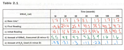
For an experiment that could measure the rate of photosynthesis, it is possible to use a dye reduction technique. In this experiment, the experimenter would measure how different frequencies of light would affect the rate of photosynthesis. Gathering a sample of plant pigments, and replacing NADP, plants natural electron acceptor, with DPIP, one could measure the rate at which photosynthesis occurs given certain conditions of the chloroplasts. As more light is absorbed and more DPIP is reduced, similarly to how NADP would be, DPIP will become colorless. This will increase the light transmission in a spectrophotometer, which will tell us the rate at which photosynthesis is occurring. To test the rate of cellular respiration, one would have to create an experiment that could possible measure the amount of oxygen that is consumed.
- Describing 2–3 different strategies that organisms employ to obtain free energy for cell processes (e.g., different metabolic rates, physiological changes, variations in reproductive and offspring-rearing strategies).
- One interesting strategy is used by animals cells during the electron transport chain. The cell will use the electronegativity of an oxygen molecule in order to cause the electrons to move up the chain towards the oxygen molecule. As the electrons move, they will release free energy to power the ETC.
- Plant cell will use the carbon dioxide that is released as a byproduct of cellular respiration in the process of photosynthesis in order to generate ATP. This carbon dioxide, alongside water and sunlight, will be used to create the sugar that is necessary for the plant cell to live.
- Refine or revise a visual representation to more accurately depict the light-dependent and light-independent (i.e., Calvin cycle) reactions of photosynthesis and the dependency of the processes in the capture and storage of free energy.
 |
| Source: www.wisegeek.com |
The picture above is very accurate in that the inputs of photosynthesis are CO2, water, and light and that the outputs are sugar and oxygen. However, it this picture were to be more accurate it would also have to depict how light energy is only used in the first phase of photosynthesis, the light reactions. It would also have to depict that from these light reactions, ATP and NADPH are produced. Then, the picture would have to show how the Calvin Cycle, the dark reactions which require no sunlight, will regenerate these two substances back into ADP and NADH as it produces the sugar the plant uses for energy, along with the byproduct oxygen. This whole process however, it driven by the capture of sunlight and the creation of free energy and ATP and is necessary for the plant to be able to live.
- Pose scientific questions about what mechanisms and structural features allow organisms to capture, store, and use free energy (e.g., autotrophs versus heterotrophs, photosynthesis, chemosynthesis, anaerobic versus aerobic respiration).
- Is there a difference between how much energy is created by plant cells during the summer or in the winter? Or in other words, is photosynthesis greatly affected by the changes in temperature during the season?
Since the pigment of the plants is found in the chloroplasts of the plant cell and does play a role in light absorption, it may be plausible to believe that as the chlorophyll breaks down, there is a lower amount of light able to be absorbed during the winter. Due to this is may be likely that there is a lower rate of photosynthetic reactions during the winter.
- Is it plausible to believe that oxygen could be replaced in the electron transport chain by another molecule?
The main feature of oxygen that is helpful to the ETC is that its high electronegativity is what draws the electrons down the chain as the oxygen molecule pulls the electrons to it. It may be plausible to believe then, that another molecule with high electronegativity may be able to cause the same reaction. However, problems may occur as the different electronegative may speed up the rate at which the electrons travel down the chain. The molecule may also cause problems in different parts of the cell and may be more difficult for the cell to acquire naturally from the environment.
- Why is Mercury a toxin to our body?
Mercury is especially detrimental towards the body because it causes a decay in the membranes of several organs, such as the liver, brain, and kidneys. It does this due to its high density, as that causes Mercury to accumulate inside of the body. Yet, Mercury is also dangerous because it causes damages to not only these organs, but also the body’s DNA and chromosomes.
 |
| Source: kids.britannica.com |
- Create a visual representation to describe the structure of cell membranes and how membrane structure leads to the establishment of electrochemical gradients and the formation of ATP.
The image below describes how the proteins that carry electrons that are embedded in the membrane serve to establish the electrochemical gradients, used especially in the Electron Transport Chain to create ATP.
 |
| This occurs in the inner membrane of the mitochondria. |
Sources:
https://thescienceinformant.wordpress.com/2011/08/25/why-is-mercury-so-poisonous/


